

In this section, we have leaflets for the Ophthalmology department. Please only read the materials on the advice of your clinician.
The leaflets are in two PDF versions:
- Booklet - for printing (double-sided, flip on short edge)
- Handout - for viewing online or downloading
The Recite Me accessibility tools can be used if you need assistance.
Advice to parents of children having dilating drops
When your child comes to the eye clinic, it is usually necessary to look at the back of their eyes and to check whether they have a refractive error (long or short sightedness).
To do this, we will need to put some dilating drops into their eyes, which will make the pupil (black part of the eye) bigger. This does not hurt your child but may sting a bit as the drops go in. He / she will need to stay still while we do this. The nurses are very experienced in putting eye drops in children’s eyes and we have different ways of doing this using toys, bubbles etc.
Aflibercept Eye Injections
Aflibercept is a medication that is injected into the eye to reduce eye complications in conditions such as diabetes, retinal vein blockage, wet age-related macular degeneration (wet AMD) and other eye conditions.
It belongs to a group of medications called anti-VEGF (Vascular Endothelial Growth Factor) agents.
Aflibercept is available under other drug names, such as Eylea, amongst others. These are all the same medication and work in the same way.
Avastin Intravitreal Injections
Your eye doctor has recommended treatment with an injection of a drug called Avastin (Bevacizumab) into your eye. This leaflet will help you to understand how the injection will help with your condition. A separate leaflet may be provided that gives you further information about your condition. You might want to discuss the information with a relative or carer.
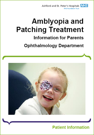
Amblyopia and Patching Treatment
Your child has a condition known as amblyopia (lazy eye) which occurs when the sight of one or both eyes is under-developed causing reduced vision.
Approximately 4% of children will need to wear a patch over one eye in order to encourage better vision in their amblyopic eye. This treatment is called patching (occlusion therapy).
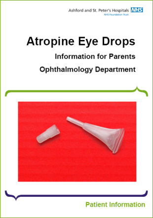
Atropine Eye Drops
Your child has had their vision checked by either a nurse or an orthoptist.
To enable us to check the health of the inside of the eye and to test for glasses accurately, the pupil (the black spot in the centre of the eye) must be dilated (enlarged.) This will be done using eye drops.
The eye drop your child will be using is called Atropine 1%.
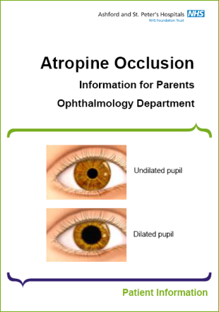
Atropine Occlusion
Your child has been diagnosed with an eye condition called amblyopia (reduced vision in one eye, sometimes known as a lazy eye).
Atropine eye drops may be used as an alternative to patching treatment. The main aim of Atropine occlusion is to temporarily blur the vision in the good eye (when viewing close objects) to promote the vision in the weaker eye.
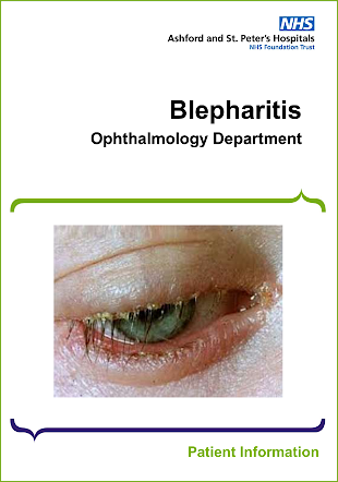
Blepharitis
Blepharitis is a common inflammatory condition of the eyelids which causes the edges of the eyelids to become red, swollen and inflamed. The most important part of managing and treating blepharitis is to keep the eyelids clean.
Botulinum Toxin for Adult Squint
Botulinum Toxin, is a chemical produced by the bacterium Clostridium Botulinum. It can be used to temporarily weaken a muscle within the body.
To move a muscle, your brain sends an electrical signal through your nerves to the muscle. This signal is passed between nerves and muscles by a chemical messenger called acetylcholine. When injected into a muscle, Botulinum Toxin temporarily stops the nerves close to the muscle from releasing acetylcholine and instructing the muscle to move, thereby the muscle is temporarily weakened.
Care of your Eye after your Injection
Today you will receive an injection into your eye. The purpose of this injection is to deliver medicine into the back of the eye to treat your eye condition and help protect your vision. Prior to the injection the nurses will check your vision and eye pressure. You will have various eye drops administered before your injection.
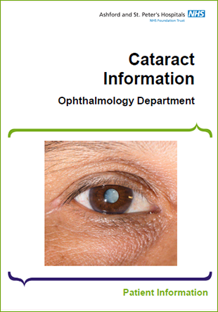
Cataract Information
A cataract is a clouding of the lens of the lens inside your eye.
Normally the lens is clear and its job is to focus light onto the back of the eye (Retina). As a cataract develops, the lens becomes opaque, and it blocks light rays passing through the lens Therefore, the vision gradually worsens.
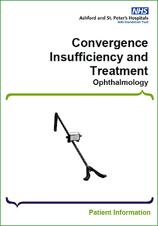
Convergence Insufficiency and Treatment
Convergence is the ability to turn both eyes inwards smoothly towards the nose when looking at a near object.
Good convergence is important for comfortable binocular vision (vision with both eyes) for any close work, including reading.
Corneal Cross-Linking (CXL)
Corneal cross-linking (CXL) is a treatment for patients with keratoconus which can prevent their condition getting worse. It is successful in more than 90% of cases. After treatment, you will still need to wear spectacles or contact lenses. Your eye will be sore for about one week after the procedure. Although vision is often hazy at first, most patients can resume contact lens wear and return to work after one week. As with all operations, there are risks: CXL is safe, but there is a small chance (about 1:30) of worse vision afterwards.
Corneal Transplantation: Deep Anterior Lamellar Keratoplasty (DALK)
The cornea is the transparent tissue at the front of the eye. It allows light to pass into the eye and focuses images so they can be seen.
Various diseases or injury can make the cornea either cloudy or out of shape. This prevents the normal passage of light so affects the vision. Corneal transplants are used to restore vision or to relieve pain in a damaged or diseased eye.
Corneal Transplantation: Endothelial Keratoplasty
The cornea is a window of transparent tissue at the front of the eyeball. It allows light to pass into the eye and provides focus so that images can be seen. Various diseases or injury can make the cornea either cloudy or out of shape. This prevents the normal passage of light through the eye and so affects the vision. Corneal transplants are used to restore vision, relieve pain or to maintain the structure in a damaged or diseased eye.
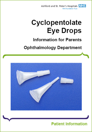
Cyclopentolate Eye Drops
Your child has had their vision checked by either a nurse or an orthoptist.
To enable us to check the health of the inside of the eye and to test accurately for glasses, the pupil (the black spot in the centre of the eye) must be dilated (enlarged.) This will be done using eye drops.
The eye drop your child will be using is called Cyclopentolate 1%.
Diabetic Retinopathy
Diabetic Retinopathy is a complication of diabetes caused by high blood sugar levels damaging the back of the eye. The light sensitive layer at the back of the eye known as the retina has a delicate network of blood vessels. These are affected by prolonged high blood sugars and become blocked or start to leak. If the retina doesn’t have a good blood supply, it cannot work properly.
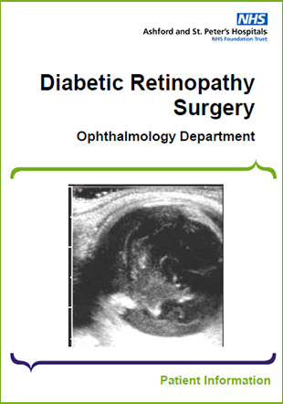
Diabetic Retinopathy Surgery
When diabetes starts to affect the retina in your eye it is called diabetic retinopathy. Diabetes can damage the retina in two main ways either causing vessels to leak (leading to swelling or diabetic macular oedema) or it can close down blood vessels.
When the blood vessels start to close down in the eye because of the diabetes, this can cause new blood vessels to grow. This is the advanced type of diabetic retinopathy (proliferative diabetic retinopathy). These new blood vessels can bleed easily and can cause bleeding into the gel of your eye (vitreous haemorrhage) which can cause floaters or reduced vision.
Digital Surveillance Clinic: AMD
As a regular patient attending the AMD (Age-related Macular Degeneration) clinic, you will have noticed how busy we are. There may have been times when you have waited a long time for an appointment and then had a long wait in the clinic.
To improve your care, we now offer a more streamlined Digital Surveillance Clinic. These clinics are a very efficient way of providing as many patients as possible with the best care available.
Digital Surveillance Clinic: Diabetic Macular Oedema
The Digital Surveillance Clinic or DSC is a fast, reliable, and effective way to follow up patients with specific eye conditions. It reduces waiting times in the clinic and ensures you have assessment and treatment (if required) within an appropriate time frame.
Digital Surveillance Clinic: Glaucoma
As a regular patient attending the glaucoma clinic, you will have noticed how busy we are. There may have been times when you have waited a long time for an appointment and then had a long wait in the clinic.
To improve your care, we now offer a more streamlined Digital Surveillance Clinic. These clinics have proved effective in other NHS hospitals and are a very efficient way of providing as many patients as possible with the best care available.
Driving and Diplopia
Diplopia means double vision. This is when you see two images of the same object at the same time. If you have diplopia, you must stop driving and inform the DVLA and your car insurance provider immediately.
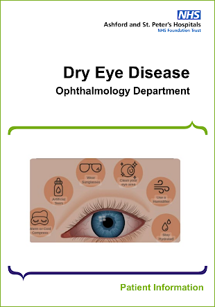
Dry Eye Disease
Our eyes are covered by a film of tears. This is to keep the surface of the eye wet, preventing damage to the eye or infection. Dry Eye Disease is where the eye does not produce enough tears, or the tears are of poor quality and evaporate off the front of the eye too quickly.
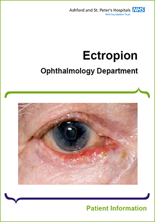
Ectropion
Ectropion is a condition where the lower eyelid turns outwards. This condition is mainly associated with age and happens due to the muscles and tissues of the eyelids becoming weaker. Entropion can also be caused by cysts, lumps or tumours of the eyelid, nerve palsies or eyelid injury or skin conditions. This can result in the eye being uncomfortable.
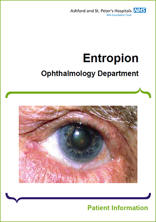
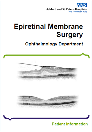
Epiretinal Membrane Surgery
An Epiretinal Membrane is a condition where a very thin layer of scar tissue forms on the surface of the retina, where the vision is sharpest. The part of the eye affected by the Epiretinal Membrane is called the Macula, which is made of special nerve cells and it provides our sharp central vision needed for seeing fine detail (reading and driving etc.)
When an Epiretinal Membrane forms over the Macula, it may contract and crumple up the Macula resulting in distorted and / or blurred vision.
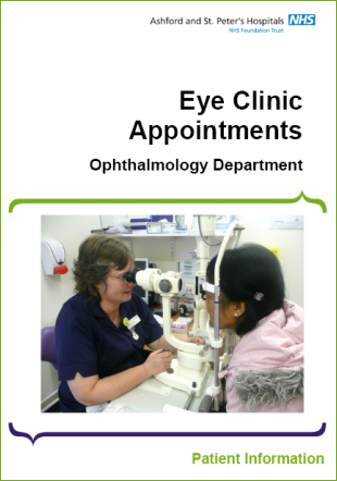
Eye Clinic Appointments
Please bring with you any distance or reading glasses you currently use. Also bring a copy of your repeat prescription or any medications you use in their original containers.
During your consultation you may need certain tests and the administration of eye drops. Please allow up to 2 hours for your appointment.
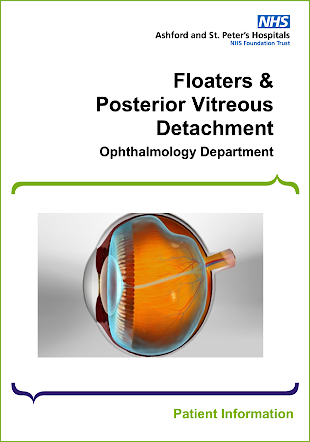
Floaters and Posterior Vitreous Detachment (PVD)
Floaters are shapes that people can see drifting across their vision. Floaters are small bits of debris floating in the vitreous gel inside the eye. They can come in a variety of forms such as small black dots, short squiggly lines or even large cobweb shapes. Shortsighted people tend to suffer from them more and they increase as we get older (a process known as vitreous syneresis). Apart from vitreous syneresis, a common cause of floaters is a posterior vitreous detachment (see below). Inflammation in the eye is a rare cause of floaters and your doctor will have examined you for this to rule this out.
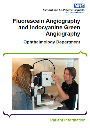
Fluorescein Angiography and Indocyanine Green Angiography
Fluorescein Angiography is referred to as FFA (Fundus Fluorescein Angiography). It is a test to look at the fine blood vessels and circulation within the retina at the back of the eye.
Indocyanine Green Angiography is referred to as ICGA. It is a test to look at the blood vessels and circulation of the choroid which is the layer below the retina in the eye.
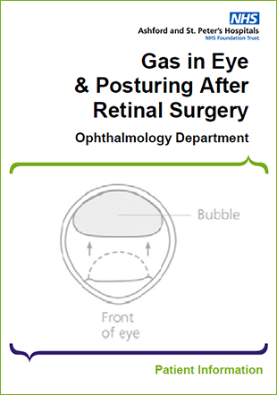
Gas in Eye and Posturing after Retinal Surgery
At the end of your eye operation your eye was filled with a medical gas. Gas is commonly used in vitrectomy eye operations to keep the retina in place whilst it is healing.
For the gas to have the right effect, it is necessary for you to keep your head in a certain position. You will have received instructions as to what your individually prescribed head posture is. This will depend on where in your retina the problem is. You must posture in such a way that the gas in your eye rises to cover the retinal problem. This is the only way for the retina to heal properly and for your sight to improve. The commonest postures are right side, left side, or face down.
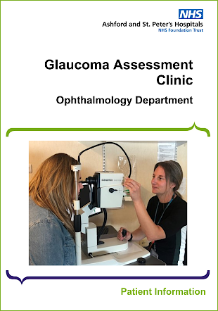
Glaucoma Assessment Clinic
The Hospital Eye Service is currently under a lot of pressure to see new patients in a timely manner, so we now offer a Glaucoma Assessment Clinic for your initial appointment with us to help reduce the time you wait to be seen for diagnosis and treatment.
These clinics have proved effective in other NHS hospitals and are a very efficient way of providing as many patients as possible with the best care available.
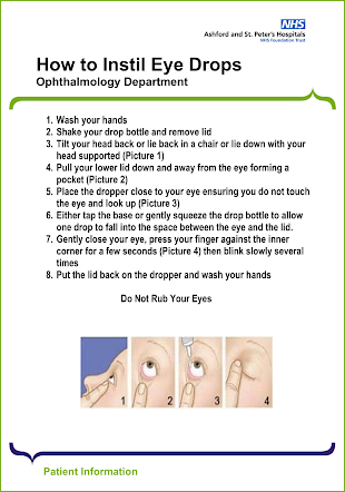
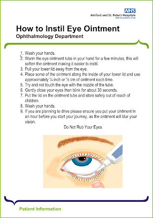
How to Instil Eye Ointment
To instil your eye ointment, please follow the steps detailed in this leaflet.
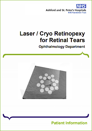
Laser / Cryo Retinopexy for Retinal Tears
Your eye doctor has told you that you have a retinal tear. This leaflet will help you to understand what has happened to your eye and the treatment that we can offer. You might want to discuss the information with a relative or carer. We will ask you to sign a consent form, so it is important that you understand the information in this leaflet before you agree to go ahead with the treatment. If you have any questions, you might want to write them down to help you remember to ask one of the hospital staff. The most common questions are listed below and will be addressed in this leaflet.
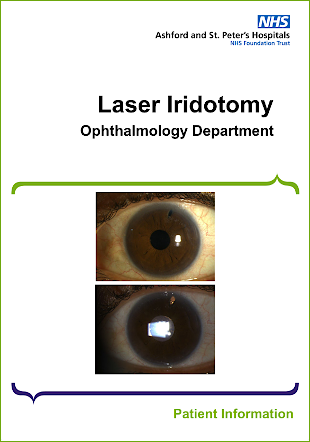
Laser Iridotomy
Laser iridotomy is a procedure whereby a laser is used to make a small hole in the periphery of the iris (coloured part of the eye).
The eye is constantly producing an internal fluid called aqueous humour and this fluid leaves the eye through the pupil and drains away through a space called the angle. This is a ring shaped, coral reef structure around the outer limit of the iris.
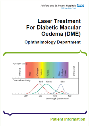
Laser Treatment For Diabetic Macular Oedema (DME)
Your specialist has informed you that you have Diabetic Macular Oedema (also called Diabetic Maculopathy). Please read this information sheet and, if you have any questions, discuss this with your specialist before you have the treatment.
Diabetes can damage small blood vessels (capillaries) in your retina. Damaged blood vessels leak fluid causing swelling of the retina. If the middle part of the retina (Macula) becomes swollen, your sight may be severely damaged.
Laser Treatment for Proliferative Diabetic Retinopathy (PDR) Pan Retinal Photocoagulation (PRP)
Your specialist has informed you that you have proliferative diabetic retinopathy and recommended laser treatment. Please read this information sheet and, if you have any questions, discuss this with your specialist before you have the treatment.
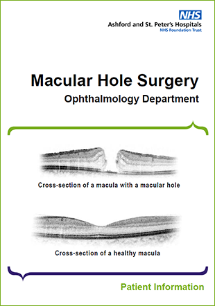
Macular Hole Surgery
The back of the eye has a light-sensitive lining called the retina, similar to the film in a camera. Light is focused through the eye onto the retina, allowing us to see. The centre part of the retina is called the macula -it is here that light must be focused for us to see fine detail, to be able to read and to see in colour.
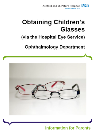
Obtaining Children's Glasses
Children are tested once a year for glasses at the hospital unless the eye care professional advises otherwise.
A voucher (HESP1) will be given to you if a new prescription is required when the doctor tests your child’s eyes for glasses.
The voucher is issued to children under 16, or under 18 and in full-time education. It is worth a fixed amount of money to help towards the cost of purchasing the glasses. The amount of money is dependent on the strength and / or type of lenses needed.
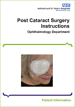
Post Cataract Surgery Instructions
Keep your eye patch on for 6 hours as instructed before you left the eye ward. After 6 hours wash your hands with soap and water, remove the entire patch and put it in rubbish bin. Do not put any part of this dressing back on.
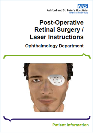
Post-Operative Retinal Surgery / Laser Instructions
Use your eye drops as prescribed and continue until instructed by the ward nurse when to stop.
Further bottles of drops can be obtained from your GP if you run out before the end.
Pre and Post-Operative Cataract Instructions
If you are having a morning operation you will ask to attend the Eye Department at 7.30am. For an afternoon appointment you will have to attend at 12.30 pm. Prior to coming for surgery please take all your regular medications unless you have been instructed by an Eye Doctor or Anaesthetist not to take them on the morning of surgery.
Ranibizumab Eye Injections
Ranibizumab is a medication that is injected into the eye to reduce eye complications in conditions such as diabetes, retinal vein blockage, wet age-related macular degeneration (wet AMD) and other eye conditions.
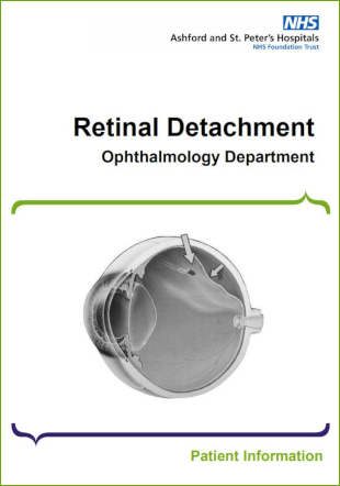
Retinal Detachment
The retina is the light sensitive film at the back of the eye and retinal detachment is a condition where the retina peels away from the inner wall of the eye. In most cases the retina detaches because a hole or a tear has formed in the retina allowing fluid to pass underneath the retina.
Most retinal detachments occur as a natural ageing process in the eye but certain people are at higher risk than others. These include people who are short sighted, those who have had cataract surgery in the past and those who have suffered a severe direct blow to the eye. Some types of retinal detachments can run in families but these are rare.
SLT Laser
Glaucoma as a disease, is the end result of a number of different conditions, which if left untreated, can cause permanent damage to the optic nerve resulting in irreversible visual loss and eventually blindness, which at the simplest level, is due to the eye pressure being too high for the state of the nerve of the eye.
Clear fluid called aqueous humour, circulates through the front of the eye providing nourishment to the tissues and helps to maintain the normal functions of the eye. The fluid contributes to the pressure in the eye, intraocular pressure (IOP), much like air in a bicycle tyre with a certain range of eye pressure being necessary for the normal functioning of the eye.
Squint Surgery for Adults
You have an eye muscle imbalance that has resulted in one of the eyes turning in, out, up or down. The squint may be present some of the time or all of the time. It can be in one eye or alternate between the eyes.
Squint Surgery for Children
A squint is one of the commonest eye conditions affecting children. It means that when a child is looking at something the two eyes are not both pointing at that object. One eye can turn in, or out, or less commonly up or down. The squint may be present some or all of the time. It can be in only one eye, or it can alternate between the eyes.
Useful Contacts for Vision After Stroke
Stroke can affect your vision in many ways. We may have given you a leaflet regarding your specific eye concern after your stroke. Here is a list of local and national services which may be of use to you regarding your vision and stroke.
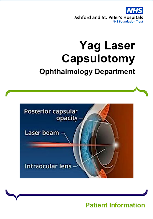
Yag Laser Capsulotomy
During your operation to remove your cataract, a very thin part of the lens called the ‘capsule’ is left behind, to act as a ‘bag’ to support the new clear lens (implant), that replaced your old lens (cataract).
This capsule or bag is very thin, like clingfilm. In most patients this capsule can thicken with time becoming opaque, this is known as a posterior capsule opacification (PCO) and gives a similar effect to having a cataract.
Your Child's Appointment in the Eye Clinic
Please allow between 2 and 3 hours for your appointment in the eye clinic. The reason for this amount of time is that your child may need to see the Orthoptist first, then have eye drops instilled and wait for these to work, before seeing the Eye Consultant.
It may be that your appointment finishes much sooner, particularly if eye drops are not required.


