


In this section, we have leaflets for the Trauma and Orthopaedics department. Please only read the materials on the advice of your clinician.
The Recite Me accessibility tools can be used if you need assistance.
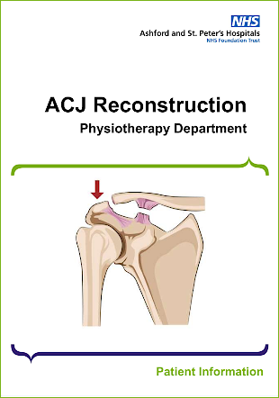
ACJ Reconstruction
The acromioclavicular joint where the collar bone (clavicle) meets the highest point of the shoulder blade (acromion of scapula). The acromioclavicular and coracoclavicular ligaments work together to hold the acromioclavicular joint in place. The acromioclavicular joint itself has little movement in isolation but supports with moving and rotating the shoulder blade (scapula).
ACL Reconstruction
The ACL connects the femur to the tibia. It functions as a stabiliser of the knee joint and it resists forward movement of the tibia on the femur and also acts to prevent the tibia from excessive rotation when pivoting.
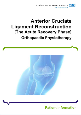
ACL Reconstruction - Acute Recovery Phase
The ACL is a broad thick band that attaches to the femur and passes forwards and downwards to attach onto the top of the tibia. The ACL has an important role in stabilising the knee during function, especially with pivoting movements and quick changes of direction.
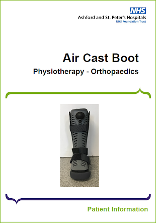
Air Cast Boot
To provide protection of your foot and / or ankle as advised by the Orthopaedic consultants. Your consultant or physiotherapist will tell you how long you will need to wear this for. Your boot will be fitted by a trained professional. You will only need to wear the boot as instructed by your therapist or doctor.
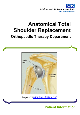
Anatomical Total Shoulder Replacement
A total shoulder replacement will involve the removal of the damaged portions of the joint. Traditionally, the head of the humerus (ball) is removed and replaced with a metal head and stem. The stem is inserted into the shaft of the long bone of the arm (the humerus) and can vary in size depending on clinical need (please see below). The socket, which is usually comprised of plastic (polyurethane) is then inserted into the glenoid (a portion of the shoulder blade).
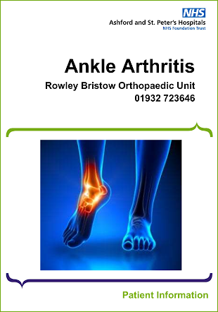
Ankle Arthritis
Ankle arthritis can cause pain, stiffness and swelling of the ankle. It occurs when the cartilage, which covers the ends of bones at joints, becomes damaged.
Cartilage usually works as a shock absorber and reduces friction during movement. When it is damaged or worn, it can lead to bone rubbing directly against bone, causing pain. Extra pieces of bone can also form around the joint, as well as scarring of the joint lining, with both processes resulting in joint stiffness and swelling.
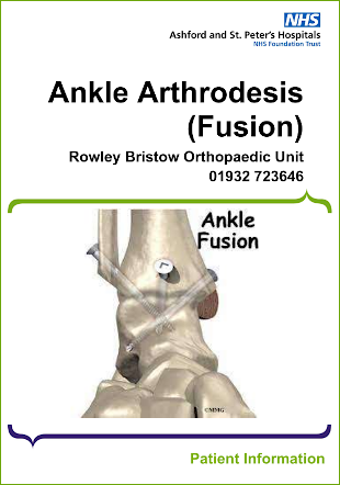
Ankle Arthrodesis
The main reason for performing an ankle fusion is arthritis. This can be age related from general wear and tear or following an ankle fracture.
Arthritis is damage to one or more joints within the body. There are two main types of arthritis, the first one, and probably the most common, is Osteoarthritis where there is a gradual wear and tear of the cartilage between the bone ends, resulting in stiffness and increasing pain. In some patients this type of arthritis can occur following an ankle fracture.
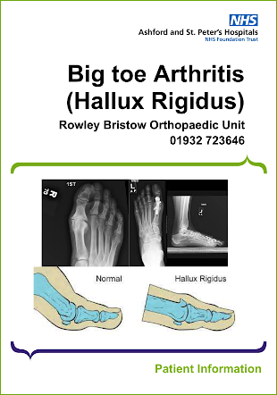
Big Toe Arthritis
Hallux rigidus is a type of arthritis in your big toe. Specifically, it affects your big toe joint - the metatarsophalangeal (MTP) joint. Your MTP joint is where the base of your big toe meets your foot.
“Arthritis” is a general term for a group of more than 100 diseases. It causes inflammation (swelling) in and around your joints. Healthcare providers sometimes call hallux rigidus “big toe arthritis.”
Hallux rigidus means “stiff big toe” - the condition’s most common symptom. It causes pain and stiffness in your MTP joint. It’s a form of osteoarthritis (“wear and tear arthritis”).
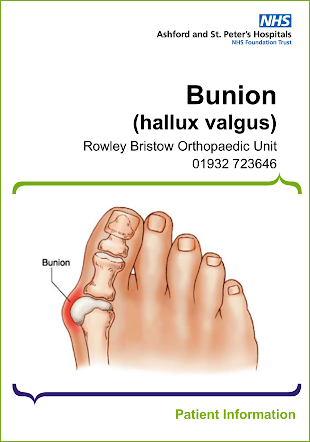
Bunion
The medical name for a bunion is hallux valgus. It is an obvious lump at the base of your big toe, caused by the joint at the base of the big toe deviating towards the second toe.
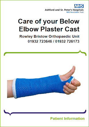
Care of your Below Elbow Plaster Cast
A backslab is usually made from plaster of Paris and does not completely encircle the whole limb. They are generally applied in Emergency departments and walk-in centres. The main reason for not applying a full plaster cast is because swelling may increase in the days after an injury. A backslab is usually temporary and will either be completed to a full plaster or changed to a new one. A full plaster encircles the limb and only allows minimal swelling.
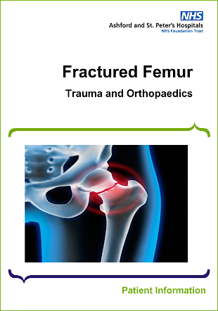
Fracture Femur
This leaflet has been designed to help you, the patient, and your relatives have a better understanding of the type of injury you have sustained and the operation you require.
During your stay in hospital if you or your next of kin have any questions, please do not hesitate to ask the ward sister and their nursing team.
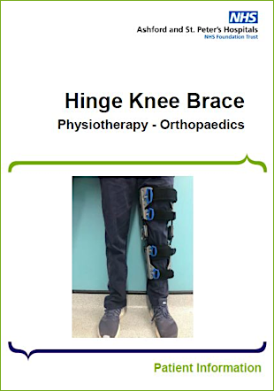
Hinge Knee Brace
Your brace will be fitted by a Physiotherapist. The fitting will be dependent on your injury type and instructions from you consultant.
Occasionally these braces slip down whilst you are walking / moving. If this occurs, unfasten the straps, reposition the brace and re-fasten to a snug fit.
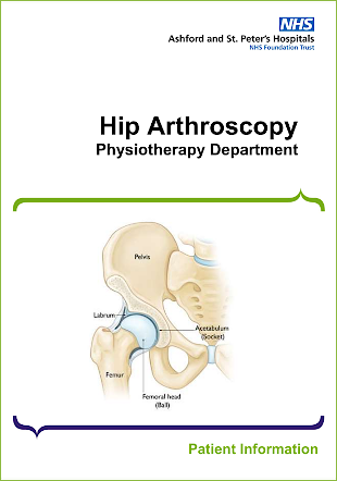
Hip Arthroscopy
Hip arthroscopy is a type of keyhole surgery used to diagnose and treat joint conditions. It is commonly performed as a day case where you are able to return home on the same day. An arthroscope with a small camera attached is inserted through a small incision over your hip, so the surgeon is able to see into your hip joint. This allows the surgeon to examine what damaged structures may need to be repaired or removed.
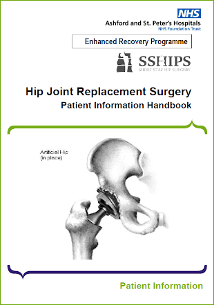
Hip Joint Replacement Surgery ERP
This booklet has been written to give you and your family / carer an understanding of a Total Hip replacement operation including things you should know before and after the operation. This is to ensure you know what to expect.
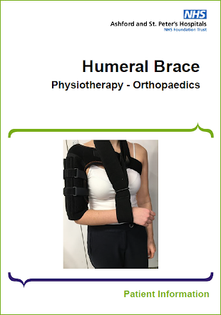
Humeral Brace
In addition to providing protection and support, the brace works by squeezing the muscles in your arm in order to keep the bones aligned. Your consultant or physiotherapist will tell you how long you need to wear the brace for and whether it can be removed for certain activities such as bathing. The brace will be sized and fitted by a nurse or physiotherapist. You may be referred to an outpatient physiotherapist to guide you through your rehabilitation and advise on further management of the brace.
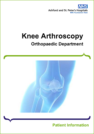
Knee Arthroscopy
The aim of arthroscopic surgery is to correct mechanical problems within your knee joint. The surgeon is able to confirm the nature of the injury and remove or repair the damaged part whilst causing minimal disruption to the knee joint. Once the structures in your joint have become damaged failure to correct the damage may result in further deterioration of your knee.
In some cases surgery will not be able to reverse the damage which has already occurred. In the majority of cases your recovery will be dictated by the degree of damage and how well you can rehabilitate your knee.
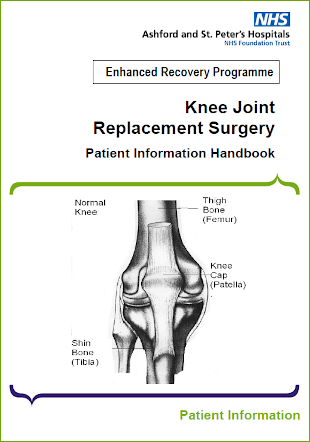
Knee Joint Replacement Surgery ERP
This booklet has been written to give you and your family / carer an understanding of a total knee replacement operation including things you should know before and after the operation. This is to ensure you know what to expect. Please note that your consultant may give you extra or slightly different advice to suit your particular case. Please ask your family / carer to read this booklet too. They will find the information helpful in understanding what your needs will be when planning your discharge.
Knee Meniscal Repair
A meniscal tear may be the result of a twist – a typical injury for someone like a netballer or footballer or from repeated squatting. These tears may cause pain, swelling and a catching or locking sensation.
If not corrected, a meniscal tear, like dirt in the ball bearings of a machine, can irritate the smooth joint surface and damage the articular cartilage. This can lead to more serious problems such as arthritis.
Knee Meniscal Root Repair
A meniscal tear may be the result of a twist – a typical injury for someone like a netballer or footballer or from repeated squatting. These tears may cause pain, swelling and a catching, locking sensation or giving way. Some are a result of degenerative changes.
A meniscal tear can happen within the meniscus itself or at its root attachment to the bone. The latter is called a root tear. Root tears are not very common and behave differently to typical meniscal tears.
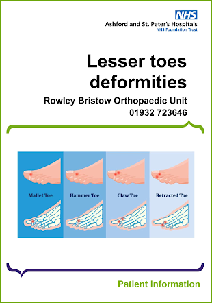
Lesser Toes Deformities
The toes which are not your big toes are known as the lesser toes. They become deformed when more pressure is exerted on them than their joints can resist. When the muscles that control the toes become unbalanced, so that one set pulls harder than others, this causes the toes to bend further. In some people the tissues in the lower part of the joint at the base of the toe (metatarso-phalangeal joint) becomes weak, allowing the base of the toe to drift upwards which further unbalances it.
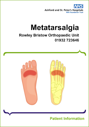
Metatarsalgia
Metatarsalgia is a general term used to refer to any painful foot condition affecting the metatarsal region of the foot. Traditionally, the term tends to refer to pain under the balls of the feet or “metatarsal heads”. However, there are a range of specific conditions that exist under the umbrella term “metatarsalgia”.
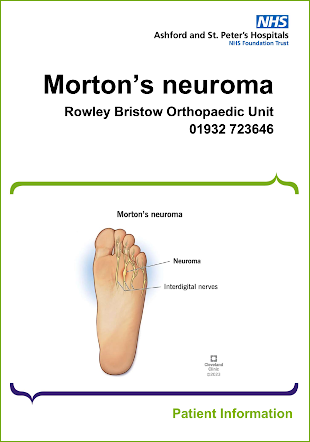
Morton's Neuroma
A Morton’s neuroma is swelling and inflammation of a nerve that lies between two metatarsal bones (toes) of your foot. It causes pain in the ball of your foot and possibly your toes. This swelling causes thickening of the nerve tissue that surrounds the digital nerve that leads to the toes. It is usually found at the base of the third and fourth toes. This condition can be exacerbated when wearing tight or high heeled shoes which can play a role in symptom cause and certainly make the symptoms worse. The lump (neuroma) is not dangerous and will not cause problems other than in the foot. Occasionally people have Morton’s neuromas in both feet.
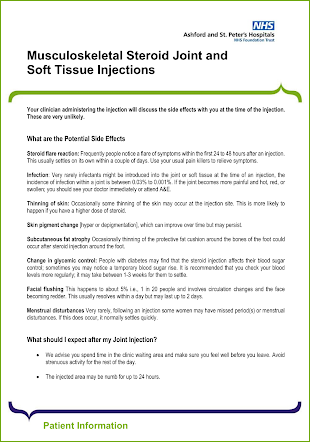
Musculoskeletal Steroid Joint and Soft Tissue Injections
Your clinician administering the injection will discuss the side effects with you at the time of the injection. These are very unlikely.
Orthopaedic Supported Discharge
The aim of Orthopaedic Supported Discharge (OSD) is to provide a greater proportion of your rehabilitation within your home setting rather than in hospital. It may not be suitable for everyone however it has been found that patients can often achieve better outcomes from rehabilitating at home rather than in a hospital setting.
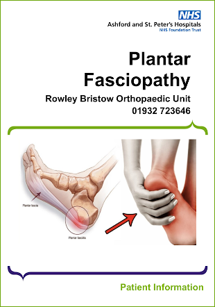
Plantar Fasciopathy
Plantar fasciopathy affects the ligaments that lie underneath the bones in the sole of your feet. The strongest ligament is the plantar fascia or aponeurosis that runs from the heel bone to the ball of the foot. This ligament is divided into three bands; the central and inside(medial) portions are the strongest. Weight bearing causes the foot to flatten, and the ligament stretches. Sometimes this ligament becomes overstretched/overloaded around the heel with time or increased activity. Changes can occur within the plantar fascia resulting in reduced elasticity and pain. The pain is usually over the heel area but can also be around the middle of the foot.
Pre-op Assessment - Information for Trauma Patients
Thank you for attending clinic today at the Rowley Bristow Unit, and for completing part of your pre-operative investigations.
Please be reminded that today’s assessment is just a part of the whole Pre-op Assessment process.
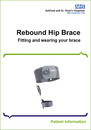
Rebound Hip Brace
The Rebound hip brace is a special orthosis, which limits bending of the hip (flexion and extension) and crossing of the leg (adduction). It is a supportive brace but is not guaranteed to prevent hip dislocation or other injury. Your doctor has determined that wearing a brace will aid in your rehabilitation. The purpose of this brace is to prevent the risk of hip dislocation. It can be used for non-operative management as well after dislocation and reduction of hip.
This leaflet will explain the things that you should do for the duration of time that you are required to wear your brace. The brace is supposed to be worn all times during walking but can be taken off whilst in bed.
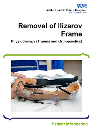
Removal of Ilizarov Frame
Depending on the site of your Ilizarov frame and how well the bone(s) are healing, the frame may be removed all at once or in stages.
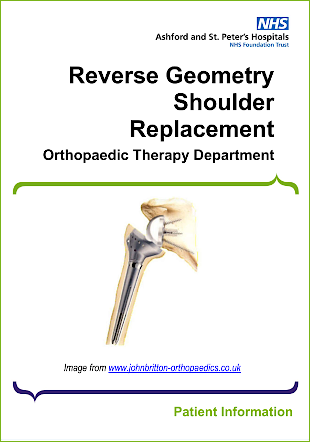
Reverse Geometry Shoulder Replacement
The Rotator Cuff is a group of four muscles that come from the shoulder blade and attach onto the ball of the ball and socket joint. It is made up of the Supraspinatus, the Infraspinatus, Subscapularis and Teres Minor (please figure 2). Their primary action is to maintain the position of the ball within the socket throughout movement. They also contribute to individual movements.
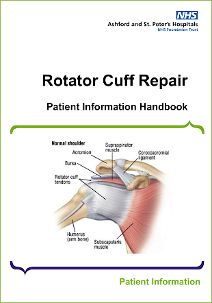
Rotator Cuff Repair
The Rotator Cuff is a group of four muscles that come from the shoulder blade and attach onto the top of the humerus (upper arm bone). It is made up of the Supraspinatus, the Infraspinatus, Subscapularis and Teres Minor. Their primary action is to maintain the position of the head of the humerus within the socket of the shoulder joint throughout movement of the arm, thus they are very important in the stability of the shoulder joint. They also contribute to individual movements.
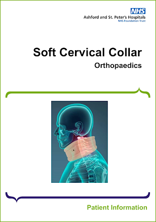
Soft Cervical Collar
A soft collar is used to protect and support your cervical spine (neck). Your doctor will have advised you wear this according to your injury. The soft collar will be fitted by a health care professional to ensure it is sized appropriately.
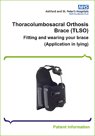
Thoracolumbosacral Orthosis Brace (TLSO) - Application in Lying
Your doctor has determined that wearing a back brace will aid in your rehabilitation. The purpose of this brace is to maintain your back in an appropriate position and limit the movement of your back to aid healing.
This leaflet will explain the things that you should do for the duration of time that you are required to wear your brace.
You are to apply your brace in LYING. Once on you are allowed to get up and mobilise. The brace must remain on until you are in the lying down position again i.e. at nighttime.
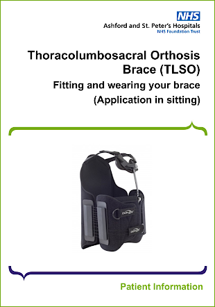
Thoracolumbosacral Orthosis Brace (TLSO) - Application in Sitting
Your doctor has determined that wearing a back brace will aid in your rehabilitation. The purpose of this brace is to maintain your back in an appropriate position and limits the movement of your back to aid healing.
This leaflet will explain the things that you should do for the duration of time that you are required to wear your brace.
You are able to apply your brace in the sitting position. It does not need to be worn in bed.
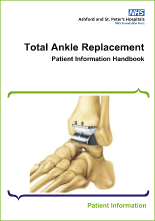
Total Ankle Replacement
In most cases, a replaced ankle will maintain the range of movement that it had prior to surgery. This means that patients can walk normally, and it is thought that the risk of putting excess strain on surrounding joints is less than would be the case with a fused ankle.
The current research suggests that, provided post-operative instructions are followed, approximately 90% of patients undergoing ankle replacement surgery can expect a significant improvement in quality of life, a reduction in pain and improved mobility. The most recent studies conclude that approximately 80 - 90% of ankle replacements will still function well 10 years after surgery.


